


Osteochondrosis of the spine is a complex of malnutrition and degenerative changes on the adjacent surfaces of the intervertebral discs and vertebral bodies, and is related to tissue destruction and structural destruction. According to the degree of injury, cervical spine, thoracic spine and lumbar spine osteochondrosis can be distinguished.
symptom
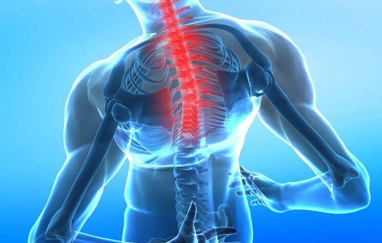
One can assume that the main sign of cervical osteochondrosis is a local change in the segmental structure of the spine (the development of lordosis, kyphosis or scoliosis)-the obvious visual curvature of the spine in the longitudinal or transverse plane. The second most common symptom is pain syndrome, which can not only be located in the vertebral area, but also in the body area innervated by the corresponding nerve roots. Another complaint of these patients is neck discomfort and fatigue.
For cervical osteochondrosis, the pain usually manifests in the neck area and can be given to the shoulder and scapula. It may be confused with the pain in myocardial infarction because it has similar symptoms. In addition, cervical osteochondrosis can be accompanied by frequent headaches and dizziness. When the arteries supplying the brain are compressed (squeezed), signs of brain dysfunction (neurological symptoms) may appear: fainting, nausea, tinnitus, mood changes, anxiety, etc.
According to the severity of the pain, it is divided into 3 degrees:
- Pain only occurs when there is significant movement of the spine;
- Relieve pain in a certain part of the spine;
- The pain is permanent.
form
According to the syndrome caused by osteochondrosis, there are:
- Compression syndrome-Compression occurs (radiculopathy-nerve root compression, myelopathy-muscle compression, neurovascular-blood vessel and nerve compression);
- Reflexes (muscle rigidity, neurodystrophy, neurovascular);
- Muscle adaptation syndrome (excessive exertion of healthy muscles while taking over the function of the affected muscles).
reason
The mechanism of the disease is the injury and displacement of the intervertebral disc due to various reasons, which leads to the loss of function of the spine devaluation (reducing pressure). The direct cause of intervertebral disc injury may be age-related degenerative changes, which are related to impaired blood supply to the intervertebral disc, mechanical damage caused by injury, and intense physical exertion of the spine-for example, being overweight.
A sedentary lifestyle plays an important role in the development of osteochondrosis, in which blood supply and intervertebral joint function are disrupted. The development mechanism of the disease is as follows: if the fibrous annulus connecting the vertebral body is damaged, the intervertebral disc is pushed back and forth-into the spinal canal cavity, or laterally-to form the median disc and the lateral disc. hernia. With the formation of a Schmorl hernia, the intervertebral disc can be pushed into the vertebral body itself-the cartilage tissue of the intervertebral disc is microscopically broken into the spongy tissue of the vertebrae. When the intervertebral disc is displaced backwards, it may compress the spinal cord and the root extending from it, which may develop into a typical pain syndrome.
diagnosis
The diagnosis of spinal osteochondrosis is based on the chief complaint, medical history data, clinical examination and instrumental examination methods. The diagnostic measure is to find out the cause of the development of neurological symptoms.
From the medical records, it can be ascertained whether there is an injury, the nature of the work-continuous physical overload (weightlifting), poor posture, work characteristics, and the position of the spine on the table and when walking, and the presence of infection.
General clinical studies (clinical blood tests, general urinalysis) and biochemical blood tests have no independent value. They are required to assess the current state, diagnose underlying diseases and emerging complications.
The diagnosis is based on the clinical manifestations of the disease and is performed by sequentially excluding diseases with similar clinical symptoms. Among the instrumental diagnostic methods, the most common and available is X-ray examination (spine imaging is a non-contrast study). It reflects the narrowing of the intervertebral joint space and allows you to identify osteophytes (bone growth) on the vertebral body, but only provides indirect information about the degree of disc damage.
Even in the early stages of the disease, an accurate diagnosis can be made through CT imaging and MRI (computational and magnetic resonance imaging) diagnosis. CT allows you to determine the smallest abnormalities in bone and cartilage tissue, MRI-performs a detailed description of the soft tissue structure and determines the location of the herniated disc.
If it is suspected that the blood supply to the brain has been violated, a duplex ultrasound scan of the cerebral arteries is performed.
Differential diagnosis of diseases with similar clinical manifestations: pathology with pain radiating to the shoulder and scapular area (liver disease, gallbladder disease, pancreatitis-pancreatitis); cervical lymphadenitis-increased cervical lymph nodes, rheumatoid arthritis; Tumor diseases (tumors of the vertebrae, roots, spinal cord and membranes), pharynx and pharynx tumors, Pancost cancer (brachial plexus compression in upper lung cancer), neck metastasis; tuberculous spondylitis-branched from tuberculosisInflammatory disease of the spine caused by bacilli; arachnoid cyst; dural pseudocyst; spinal abnormality; fibromyalgia is a disease that causes pain in muscles, ligaments, and tendons. Disease syndrome and shoulder girdle caused by excessive pressure on the neurovascular bundles between the muscles and the middle scalene muscle, above the first rib and below the clavicle, myofascial neck muscles-chronic pathology caused by local muscle spasm or seal formationSituation, represented by pain points.
Main laboratory tests used:
- Clinical blood test;
- Blood chemistry.
Main instrument research used:
- X-rays of the spine (chirograph);
- Magnetic resonance imaging (MRI);
- Computer tomography (CT);
- Perform an ultrasound duplex scan of the cerebral arteries (if insufficient blood supply to the brain is suspected).
Other instrument research used:
- Densitometry-measurement of bone density (according to indication).
treatment
The treatment of osteochondrosis of the spine depends entirely on the stage and extent of the development of osteochondrosis. In the initial stage, preventive measures, physical therapy exercises, simulator exercises and fitness can be used. For severe pain syndromes, patients need physical rest. Prescription anti-inflammatory and antispasmodic drugs. When the pain causes muscle spasm and the intervertebral disc is more intensely compressed, paravertebral block can be performed with anesthetics to open up the pathological circulation, thereby increasing the pain itself.
Apply warm ointment locally to the skin of the vertebral area to improve local blood supply and reduce tissue edema. These patients wore corsets. In patients in the initial stage of the development of osteochondrosis, chondroprotective agents are effective-drugs that improve cartilage tissue recovery, as well as drugs that improve local blood supply, venotonics, B vitamins. In the case that the pain syndrome has not stopped, there has been a long-term clinical practice of intervertebral disc root compression and spinal cord, showing that the damaged intervertebral disc was surgically removed. In cases where the intervertebral disc completely compresses the spinal cord, early surgery is required.
You should not wait until a person starts urinating or defecation spontaneously-in this case, the damage to the spinal cord may be irreversible. As physical therapy procedures, magnetic therapy, ultrasound, massage, manual therapy, acupuncture and physical therapy exercises are prescribed.
complication
Possible vegetative vascular dystonia and heart damage, cerebrovascular accidents, hypotension and hypertension (reduced and elevated blood pressure), vestibular dysfunction (impaired motor coordination), vertebral artery syndrome (narrowing of the vertebral artery)Caused by disease), periarticular disease (a disease in which mobility is impaired) of the shoulder joint.
prevention
In order to prevent osteochondrosis, it is necessary to deal with the factors that cause it, namely: avoiding spinal injuries, spinal pressure (weightlifting), and fighting overweight. For people who have suffered from the early stages of osteochondrosis, it is recommended to wear a corset at home and during physical activities. In order to let the spine rest during sleep, it is recommended to sleep on orthopedic mattresses and pillows.
What questions should you ask your doctor
Is there any exercise to help relieve symptoms?
Which drugs help treat cervical osteochondrosis?
What happens if you don’t start treating the disease in time?
Patient advice
Exercising, losing weight if you are overweight, using cold or hot compresses can help alleviate the symptoms of thoracic osteochondrosis. It is also important to eat correctly, monitor the spine, treat chronic diseases, and avoid injuries.